


Pregnancy ultrasound diagnostics - Prenatal ultrasound in Warsaw
See also: Prenatal Tests.
Modern ultrasound device
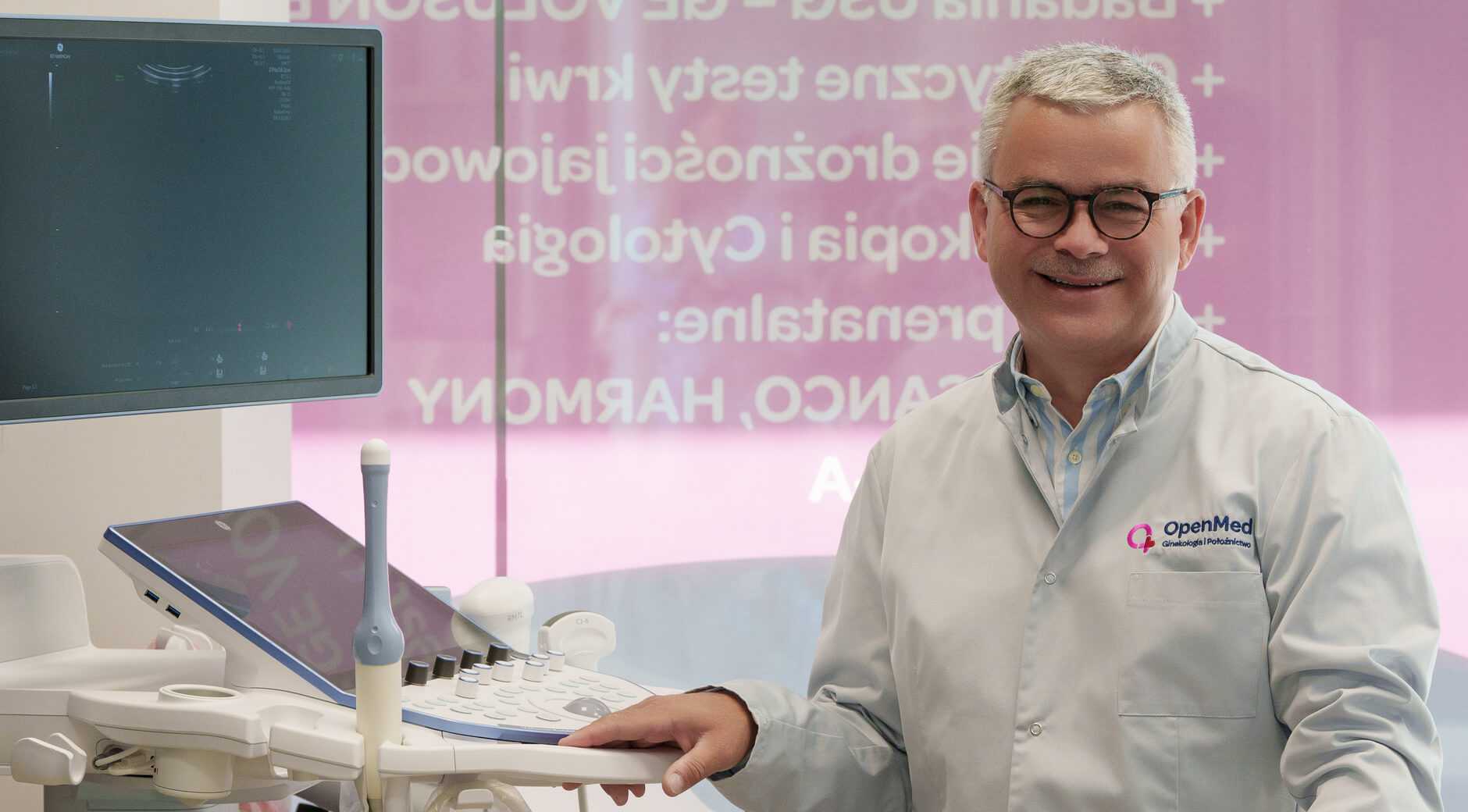
We use the GE VOLUSON Expert 22 device for ultrasound examinations.
OpenMed Gynecology and Obstetrics proudly announces that we have the latest GE VOLUSON E22 ultrasound device – one of only three devices of this class available in Mazovia. This device provides:
-
Exceptional accuracy and image quality
-
Faster and more comfortable examinations
-
Safe and painless examinations
Patient monitor during examination
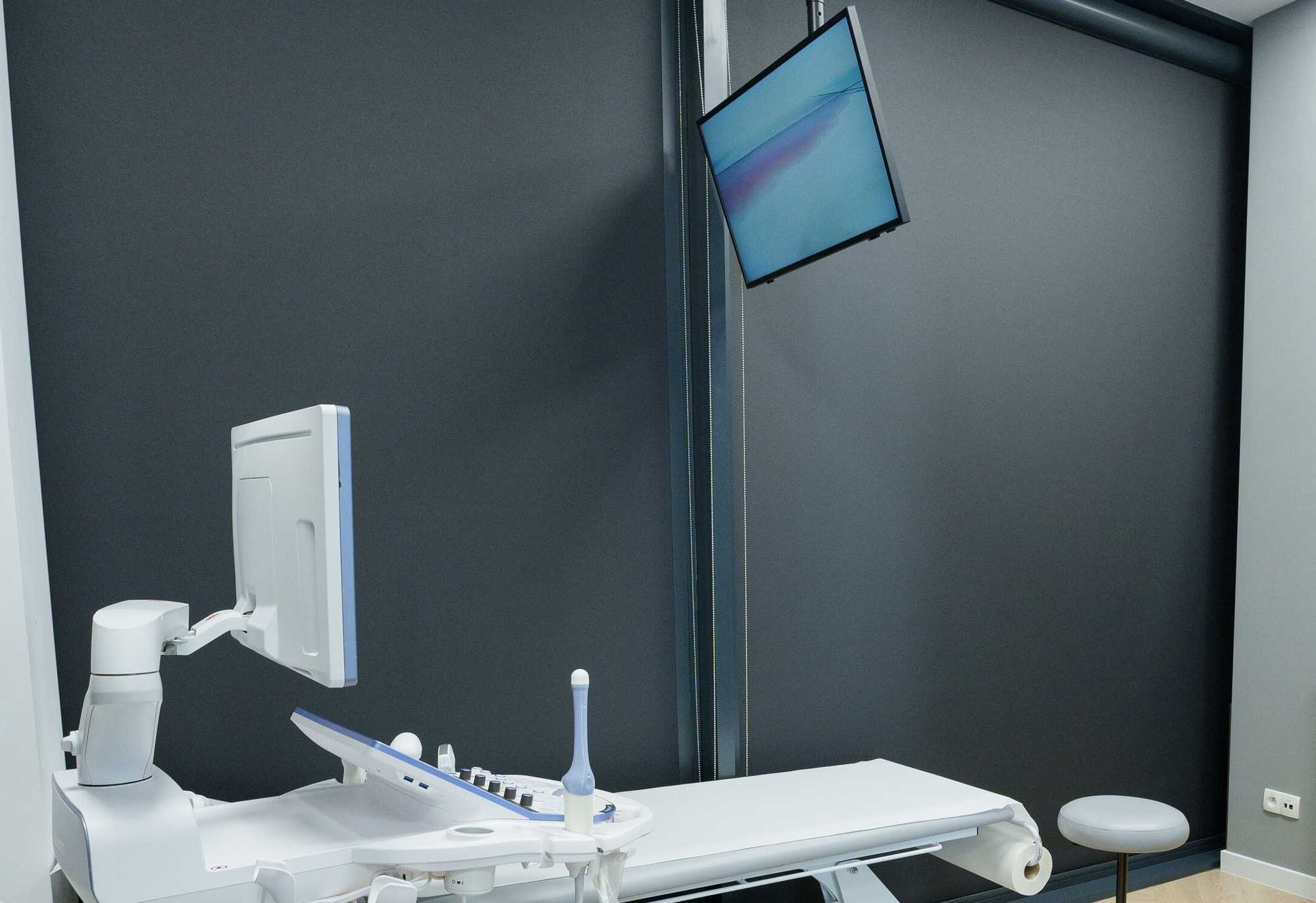
During the ultrasound examination, we provide the opportunity to observe high-quality images on a monitor mounted under the ceiling directly above the bed, allowing the patient to actively participate in the diagnostics.
3D and 4D ultrasound examination
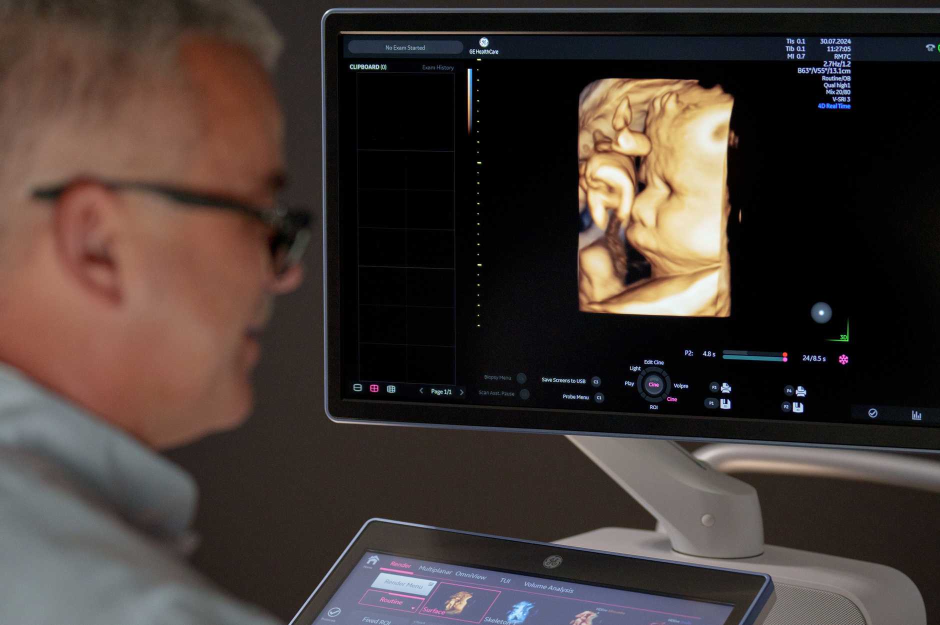
At OpenMed Gynecology and Obstetrics, we offer modern 3D and 4D ultrasound examinations that provide an exceptionally detailed image of your little one. We invite you to schedule an appointment and take advantage of advanced diagnostic technologies!
Ultrasound up to 10 weeks of pregnancy
At this stage of pregnancy, we focus on:
- Verification of the presence and position of the fertilized cell,
- Checking the gestational sac in terms of its size and shape,
- Assessment of the existence of the embryo, its heart activity, and estimated gestational age,
- Determining the number of embryos and structures such as chorions and amnions,
- Analysis of the yolk sac,
- Assessment of the female reproductive organ, including the structure, size, and shape of the uterus and adnexa.
First trimester ultrasound
This examination is recommended to be performed between 11 and 13+6 weeks of pregnancy. It focuses on the analysis of fetal anatomy and identification of genetic risk markers, which are crucial for assessing the possibility of genetic defects in the child. Precision and compliance with standards are key here.
The examination includes:
- Assessment of fetal heart activity,
- Measurement of crown-rump length of the fetus,
- Detailed analysis of fetal anatomy, including head, abdomen, heart, spine, limbs,
- Assessment of chromosomal defect markers, such as nuchal translucency, nasal bone, ductus venosus flow, and tricuspid valve flow.
Second trimester ultrasound
Scheduled for the period between 18 and 24 weeks of pregnancy, with the optimal time between 20 and 22 weeks. This examination enables accurate assessment of fetal position, its weight, anatomy, growth, as well as the condition of the placenta, umbilical cord, amount of amniotic fluid, and heart function.
Third trimester ultrasound
Performed between 28 and 32 weeks of pregnancy, most commonly between 30 and 32 weeks. The goal is to assess the same parameters as in the second trimester examination, with emphasis on pregnancy development and fetal well-being.
Pre-delivery ultrasound
Can be performed in the perinatal period, between 36 and 40 weeks, to assess fetal well-being, risks threatening the child, fetal anatomy, position, vascular flows, and child activity, as well as its body weight, which can be helpful in planning delivery.
Fetal echocardiography (fetal heart echo)
The examination includes assessment of the chest anatomy, fetal heart, and its function in case of suspected abnormalities. It is most commonly performed in the second trimester of pregnancy, but when there are indications for expanded diagnostics, the examination can also be performed in the third trimester.
Information obtained during fetal examination can be helpful in making decisions about the further course of pregnancy and the place and time of delivery. Children with diagnosed heart defects should be born in specialized centers with appropriate care conditions. This examination is also recommended when defects of this organ occur in the family.
The examination is non-invasive, painless, and completely safe. Duration depends on conditions during the examination.
Do you have any questions? Call us - 22 100 45 20.
You can check the cost of pregnancy ultrasound diagnostics and other services in our price list.